


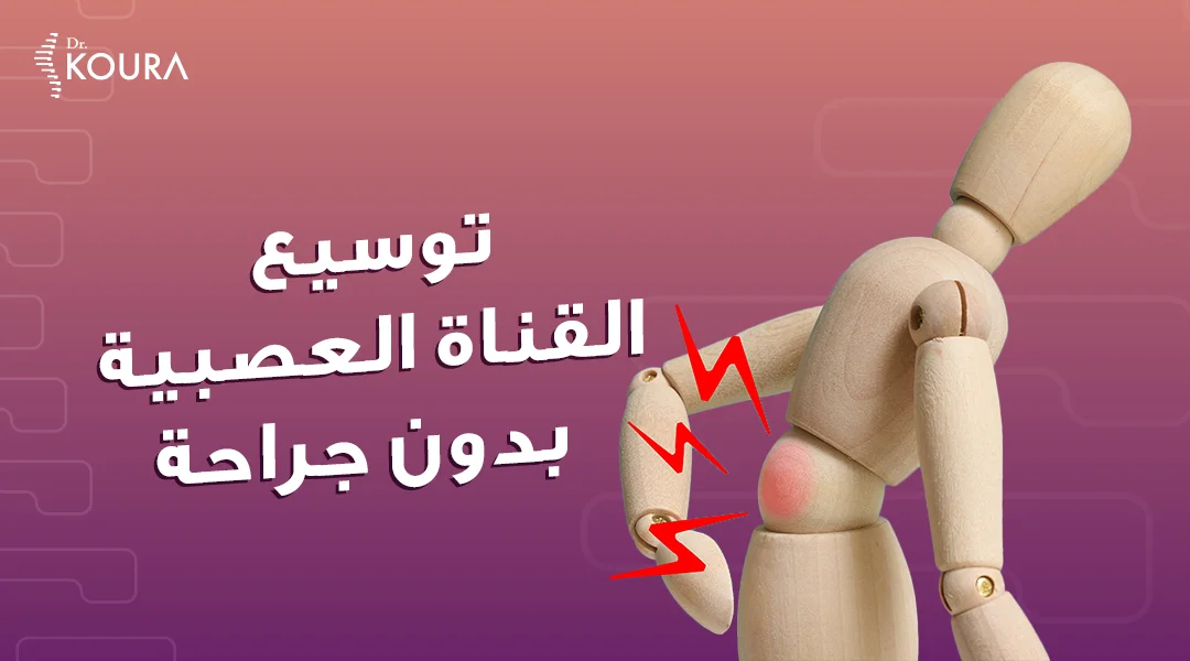
Treatment of spinal canal stenosis without surgery
Neural Canal Decompression Without Surgery and Its Advantages
The neural canal is a vital component in the human body, playing a crucial role in transmitting neural signals and information between various organs. However, this canal can encounter some issues, one of which is neural canal stenosis that can lead to various symptoms such as lower back pain, numbness, tingling, and more. Nevertheless, there is no need to worry, as with significant advancements in techniques and therapeutic methods, it is now possible to widen the neural canal without surgery.
In the following paragraphs, we will explain what neural canal stenosis is, its symptoms, causes, and complications. Additionally, we will clarify the treatment methods and the advantages of non-surgical treatment.
What is Neural canal Stenosis?
Neural canal stenosis is the narrowing of one or more spaces within the neural canal. The neural canal is the tunnel that passes through each vertebra in the spine. The narrowing of the space inside the neural canal can lead to the constriction of the spinal cord and the nerves branching out from it (nerve roots).
The narrowed space may result in the irritation, compression, or pinching of the spinal cord or nerves.
What are the symptoms of Neural canal Stenosis?
Symptoms of neural canal stenosis vary from person to person depending on the severity of the condition and the areas where pressure on the spinal cord affects nerves and nerve roots. These symptoms may include:
- Lower back Pain.
- Pain may start in the buttocks and extend down the leg, potentially reaching the foot.
- Numbness or Tingling ("Pins and Needles") may be felt in the buttocks, leg, or foot.
- Weakness may occur in the arms or legs, and it can affect one or both sides.
- Pain tends to worsen when standing for extended periods, walking, or walking on an incline.
- Pain may disrupt sleep, with episodes of electric shock-like sensations causing a burning feeling in the affected limbs.
- Neurogenic Claudication: This is a condition where the patient experiences contraction and heaviness in leg muscles. The sensation is often described as a heaviness hanging from the patient's foot, compelling them to sit down. This condition typically improves when squatting or leaning forward, leading the individual to believe it's just muscle strain, but in fact, it is not.
What are the causes of Neural canal Stenosis?
There are several reasons that may lead to neural canal stenosis, including:
- Genetic Factors involve enlargement of spinal vertebrae or tendons, causing pressure on the neural canal.
- Vertebral Slippage that causes narrowing from various directions.
- Spinal Degeneration leads to the formation of prominent bony outgrowths that compress the canal from different angles.
- Herniated Disc presses on nerves and causes frontal neural canal stenosis.
- Enlarged Posterior Ligaments that may lead to narrowing the neural canal from the back.
What are the complications of Neural canal Stenosis?
Neglecting the treatment of neural canal stenosis can lead to various complications, including:
- Loss of Bladder Control (Urinary Incontinence).
- Sexual Dysfunction because of Neurological issues, such as erectile dysfunction.
- In Rare Cases, Neural canal stenosis can lead to partial or complete paralysis in the legs.
What are the treatments of Neural canal Stenosis?
There are several methods that can be used to treat neural canal stenosis, including:
Medications
The doctor may prescribe certain medications to treat neural canal stenosis in the early and moderate stages, relying on pain relievers and anti-inflammatory drugs to alleviate pain.
Physical Therapy
The doctor may design a tailored exercise program for the patient to improve balance, flexibility, and spinal stability, while strengthening the back and abdominal muscles.
This should be supervised by a medical professional to avoid incorrect movements that could exacerbate the problem rather than resolve it. This method can be effective in the early and moderate stages to a certain extent.
Surgery
Surgery is considered a solution in advanced cases, but it is not the ideal option for all cases. Non-surgical techniques may have a role in some cases.
Non-surgical treatment for neural canal stenosis
Non-surgical treatment for neural canal stenosis is considered one of the best therapeutic approaches due to its numerous advantages compared to other methods. These include:
- Does not cause any side effects on organ functions, such as the liver and kidneys, as seen with medication-based treatments.
- Does not require major surgery, leading to a shorter recovery time.
- Rapid and evident effects, in contrast to traditional physical therapy.
- One of the key advantages of non-surgical treatment is the ability to leave the hospital on the same day and return to normal life activities the following day.
- Non-surgical treatment for neural canal stenosis involves making a small incision in the skin to introduce tools for widening the neural canal. This is done following the injection of a contrast dye into the canal, which accurately reveals the details of the neural canal and its branches. These tools help in dilating and opening the narrow areas in the neural canal, relieving pressure on the nerves and the arteries that supply them. This ensures sufficient and proper blood flow, leading to immediate improvement for the patient.
Procedures of non-surgical neural canal decompression:
- Caudal epidural injection using navigation catheter.
- Epiduroscopy, especially after neural canal stenosis caused by adhesions resulting from failed back surgry.
Can Neural canal stenosis be prevented?
Neural canal stenosis cannot be entirely prevented, but there are certain measures that can help maintain spinal health and reduce the risk of developing stenosis. These measures include:
- Consuming a Healthy Diet, and be sure that you get enough calcium in your diet to maintain bone strength.
- Maintaining a Healthy Weight:
- Keep a healthy weight to reduce stress on the spine.
- Quitting Smoking, as smoking damages arteries, contributing to back pain and making it harder for injuries to heal.
- Sitting with Proper Posture.
- Engage in regular physical activity to maintain muscle strength, especially in the back muscles.
In conclusion, having learned about neural canal stenosis, its symptoms, complications, and non-surgical treatment approaches, there is no need for concern. At Dr. Mohammad Koura's center, we offer non-surgical interventions to get rid of chronic pain issues without surgery. Therefore, it is advisable to visit the center for a prompt diagnosis and determination of the appropriate non-surgical treatment method for you.
Book with us at the following numbers for the best medical service.
 Egypt
Egypt